Explore the Microscopic World
Hands-on microscopy guides, stunning specimen close-ups, and honest gear reviews — written from real observation, trusted by students, hobbyists, and educators.
🧭 Browse by topic
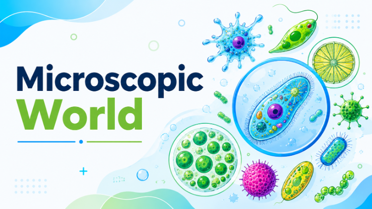
🦠 Microscopic World
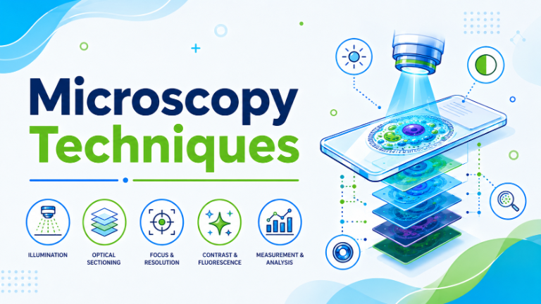
💡 Microscopy Techniques
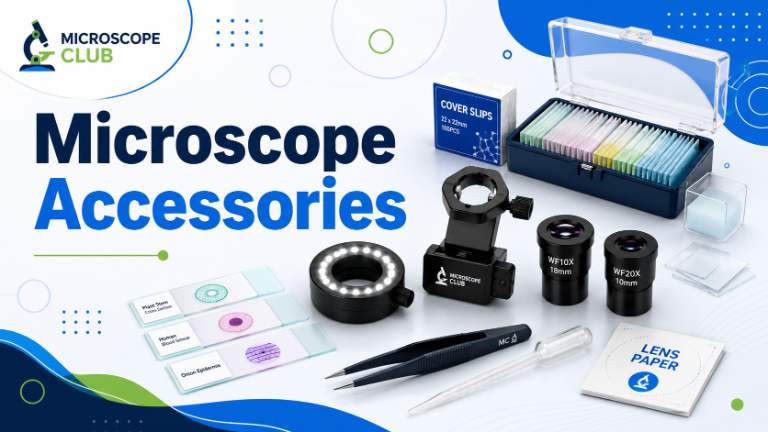
🧰 Microscope Accessories
🚀 New to microscopy? Start here
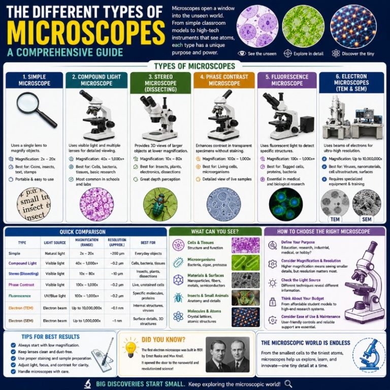
The Different Types of Microscopes
From light to electron, stereo to digital — there’s a microscope for every job. This guide walks you through every major type, how each one works, and which is the right fit for what you want to see.
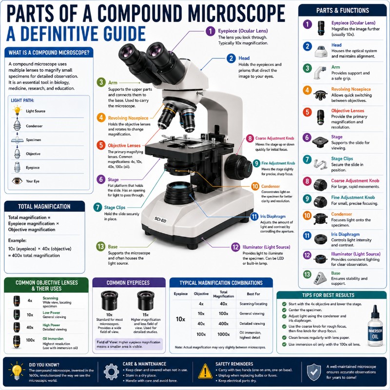
Parts of a Compound Microscope
Eyepiece, objectives, condenser, stage — a compound microscope has a dozen working parts. Learn exactly what each one does and how they work together, so nothing on your scope is a mystery again.
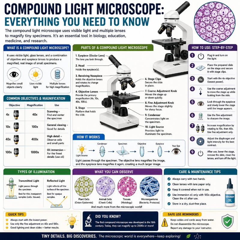
Compound Light Microscope
The workhorse of classrooms and home labs. Understand how a compound light microscope magnifies, what it can and can’t reveal, and the simple habits that get you a sharp, bright image every single time.
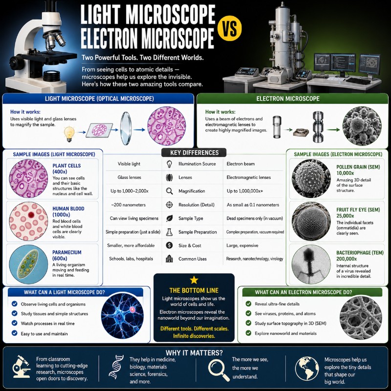
Light vs Electron Microscope
What’s the real difference — and does it matter for you? A clear breakdown of how light and electron microscopes compare on magnification, resolution, cost, and what each one can actually show you.
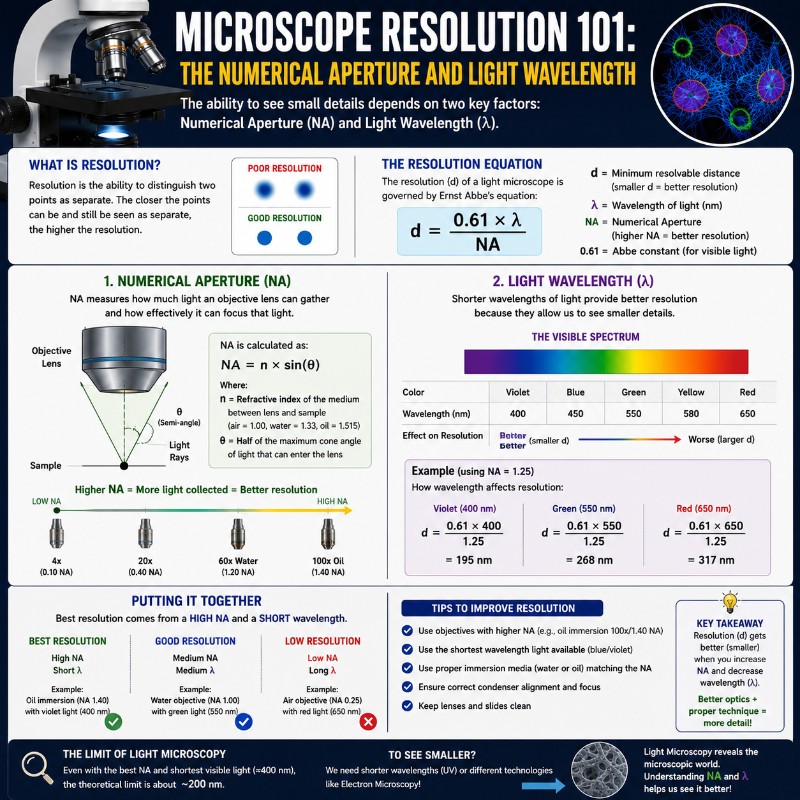
Microscope Resolution Explained
Resolution is why two microscopes at the same magnification can look completely different. Learn what limits it, what numerical aperture actually means, and how to get the sharpest image your scope can produce.
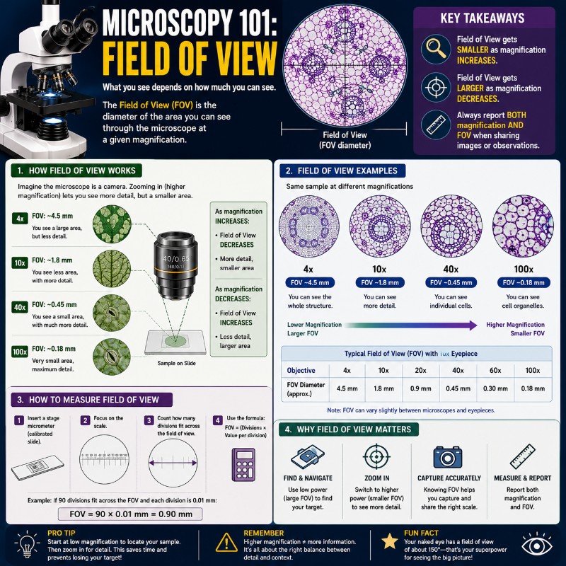
Field of View 101
As magnification goes up, your field of view shrinks — and beginners are often surprised by how fast. Understand what field of view means, how to calculate it, and why it changes everything about finding a specimen.
🔥 Most popular guides
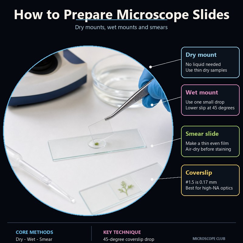
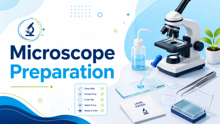
How to Prepare Microscope Slides
Wet mount, dry mount, and stained slides explained step by step — with the small techniques that stop bubbles, blur, and crushed specimens ruining your view.
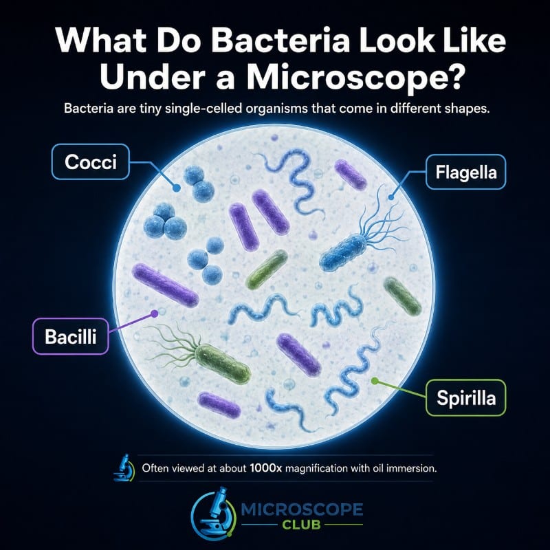
Bacteria Under the Microscope
What bacteria actually look like at high magnification, the staining that makes them visible, and how to spot the main shapes — rods, spheres, and spirals — on your own slides.
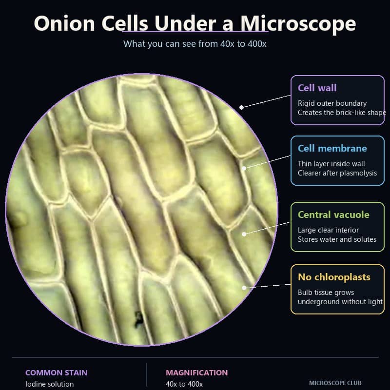
Onion Cells Under the Microscope
The classic first experiment. See the neat brick-wall pattern of plant cells, find the nucleus, and learn the iodine trick that makes everything pop into focus.
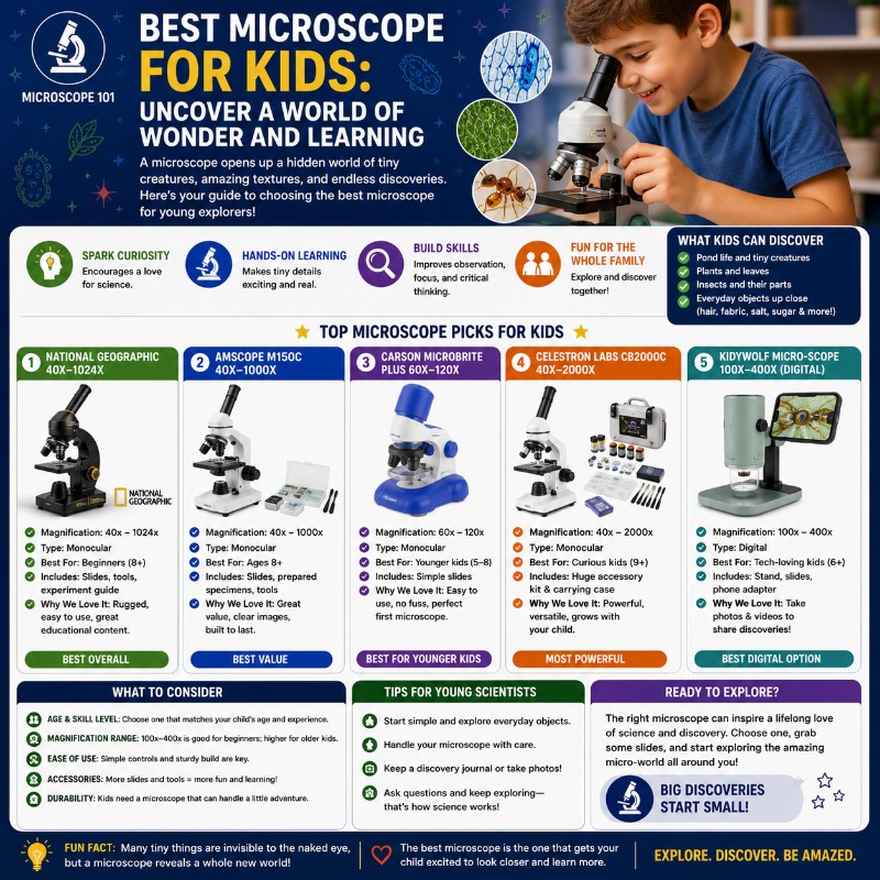
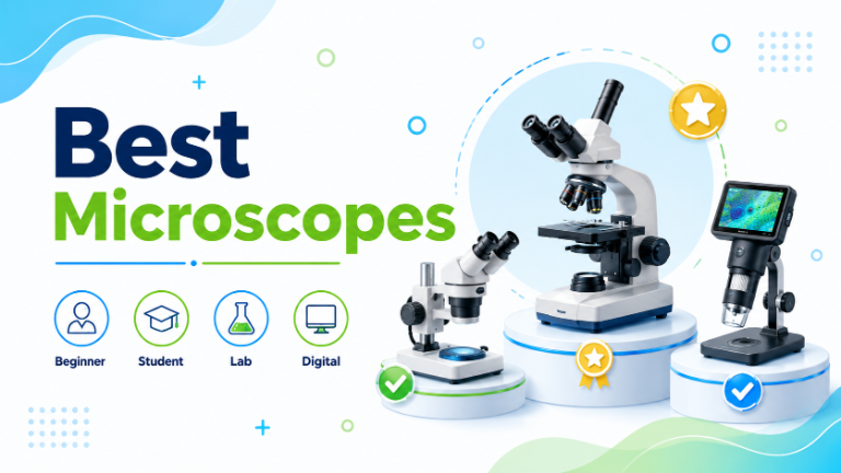
Best Microscopes for Kids
Durable, easy, genuinely fun microscopes that survive young scientists — picked by age and budget, with what to look for so you don’t waste money on a toy that disappoints.
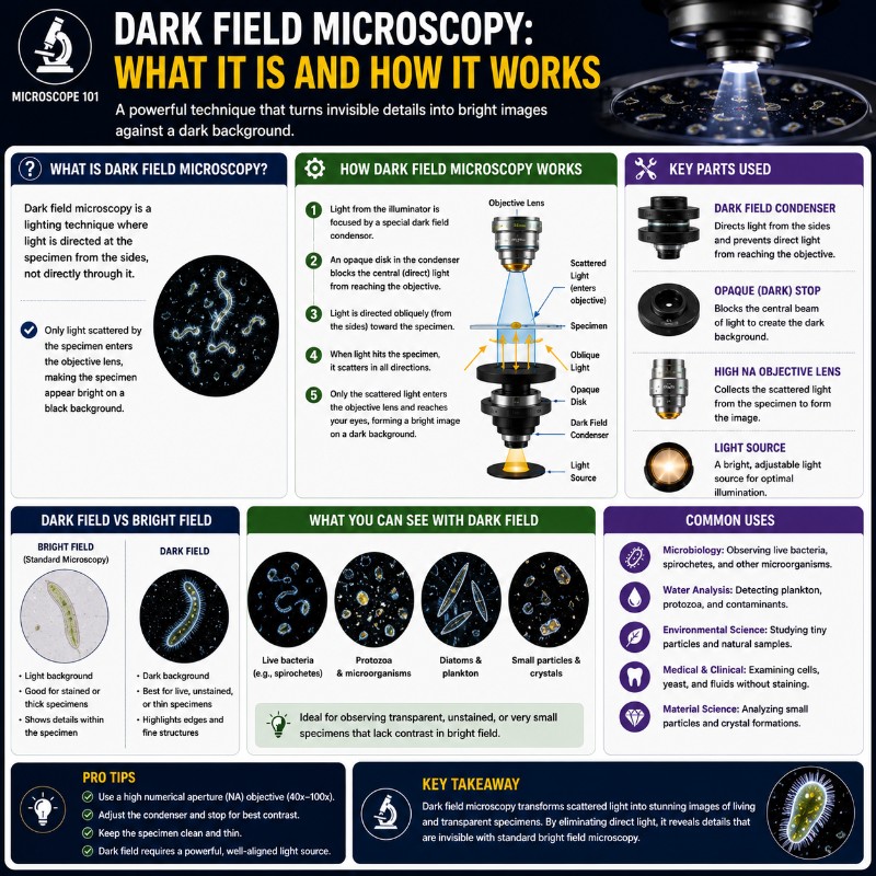
Dark Field Microscopy
The technique that makes transparent, living specimens glow bright against a black background. How it works, when to use it, and how to set it up on a standard scope.
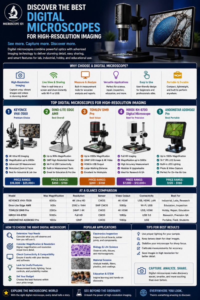
Best Digital Microscopes
Capture and share what you see. The best digital microscopes for photos and video, compared across resolution, magnification, and price for every kind of user.